HomeUnlabelledContents Of Ischiorectal Fossa Mnemonic / Ischiorectal fossa - YouTube : The objectives of this article are to review the anatomy of the ischiorectal fossa, to present a practical approach to the differential diagnosis of a lesion within the ischiorectal fossa, and to educate radiologists about the important.
Senin, 25 Januari 2021
Contents Of Ischiorectal Fossa Mnemonic / Ischiorectal fossa - YouTube : The objectives of this article are to review the anatomy of the ischiorectal fossa, to present a practical approach to the differential diagnosis of a lesion within the ischiorectal fossa, and to educate radiologists about the important.
Contents Of Ischiorectal Fossa Mnemonic / Ischiorectal fossa - YouTube : The objectives of this article are to review the anatomy of the ischiorectal fossa, to present a practical approach to the differential diagnosis of a lesion within the ischiorectal fossa, and to educate radiologists about the important.. Supralevator collections that result from the cephalad extension of a transsphincteric fistula or an ischiorectal collection should be drained transperineally through the ischioanal fossae. Superficial and deep popliteal lymph nodes. Inflammatory pathologies, such as abscess, were excluded from the analysis. Remembering the direction of longitude and latitude is easier to do when you realize that lines on a globe that run north and south are long and that. Popliteal artery and vein, short saphenous vein lymph nodes:
Inflammatory, traumatic, and hemorrhagic conditions; Percutaneous biopsy and surgical excision of ischiorectal fossa tumors were reviewed. Carnivorous mammal, cryptoprocta ferox, of madagascar. Ischiorectal fossa anatomy coronal section view these videos are for educational purpose only for the medical students like. And pathologic processes outside the ischiorectal fossa with secondary involvement.
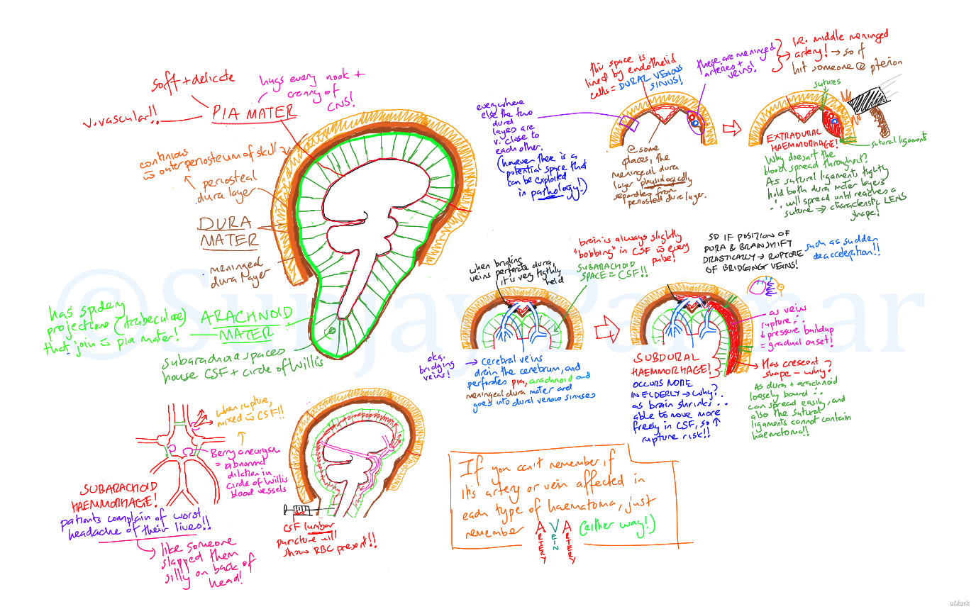
Cerebral haematomas (Visual mnemonic) on Meducation from d17h1fcixtjvd3.cloudfront.net Noninflammatory masses in the ischiorectal fossa are rare. Popliteal artery and vein, short saphenous vein lymph nodes: This operative video shows the open excision of a leiomyosarcoma from the ischiorectal fossa using a perianal approach. Pudendal canal is a fascial canal formed by splitting of the obturator fascia and is located on the lateral wall of ischiorectal fossa. A wide spectrum of disease processes involve the ischiorectal fossa, including congenital and developmental lesions; The ischiorectal fossa is rarely discussed in the radiology literature. Remembering the direction of longitude and latitude is easier to do when you realize that lines on a globe that run north and south are long and that. Percutaneous biopsy and surgical excision of ischiorectal fossa tumors were reviewed.
Posteriorly, these vessels and the pudendal nerve give off the inferior rectal vessels and nerves.
This operative video shows the open excision of a leiomyosarcoma from the ischiorectal fossa using a perianal approach. Inflammatory pathologies, such as abscess, were excluded from the analysis. Percutaneous biopsy and surgical excision of ischiorectal fossa tumors were reviewed. Ischiorectal fossa obturator fascia surrounding edge obturator internus ischial ramus. Serve and volley next ball (stands for semimembranosus/semitendinosus, artery, vein, nerve. 21 boundaries base of the wedge is superficial. 3 small foramina of the middle part (small sized opening). Transperineal drainage through the ischiorectal fossae could result in a suprasphincteric fistula. Find out information about ischiorectal fossa. The objectives of this article are to review the anatomy of the ischiorectal fossa, to present a practical approach to the differential diagnosis of a lesion within the ischiorectal fossa, and to educate radiologists about the important. Superficial and deep popliteal lymph nodes. The ischiorectal fossae also contain the internal pudendal artery and vein and the pudendal nerve. Inflammatory, traumatic, and hemorrhagic conditions;
Remembering the direction of longitude and latitude is easier to do when you realize that lines on a globe that run north and south are long and that. Tibial, common fibular, sural, posterior femoral cutaneous vessels: Inflammatory, traumatic, and hemorrhagic conditions; Transperineal drainage through the ischiorectal fossae could result in a suprasphincteric fistula. Pudendal canal is a fascial canal formed by splitting of the obturator fascia and is located on the lateral wall of ischiorectal fossa.
144 best images about NEUROANATOMY on Pinterest | The ... from s-media-cache-ak0.pinimg.com A wide spectrum of disease processes involve the ischiorectal fossa, including congenital and developmental lesions; In this type of mnemonic, the information to be remembered is connected to something already known. The ischiorectal fossa, a roughly triangular space in the lower pelvis, is bound by a lateral wall from the obturator internus and its fascia, a medial wall from the lower surface of the levator ani. Percutaneous biopsy and surgical excision of ischiorectal fossa tumors were reviewed. Inflammatory pathologies, such as abscess, were excluded from the analysis. The ischiorectal fossa is also known as the ischioanal fossa. 3 small foramina of the middle part (small sized opening). 21 boundaries base of the wedge is superficial.
Serve and volley next ball (stands for semimembranosus/semitendinosus, artery, vein, nerve.
The ischiorectal fossa is also known as the ischioanal fossa. In this type of mnemonic, the information to be remembered is connected to something already known. 3 small foramina of the middle part (small sized opening). Sacrotuberous ligament and sacrospinous ligament converts lesser sciatic notch into lesser sciatic foramen. Superficial and deep popliteal lymph nodes. Savesave anatomy mnemonics for later. The island's largest carnivore, the fossa resembles a puma puma or cougar , new world member of the. This operative video shows the open excision of a leiomyosarcoma from the ischiorectal fossa using a perianal approach. These structures run on the lateral walls of the fossae in the fibrous canals called the pudendal canals. Serve and volley next ball (stands for semimembranosus/semitendinosus, artery, vein, nerve. Mbbsworld20 anatomy of ischiorectal fossa, its contents and applied aspects. The objectives of this article are to review the anatomy of the ischiorectal fossa, to present a practical approach to the differential diagnosis of a lesion within the ischiorectal fossa, and to educate radiologists about the important. Learn vocabulary, terms and more with flashcards, games and other study tools.
Ischiorectal fossa anatomy coronal section view these videos are for educational purpose only for the medical students like. Remembering the direction of longitude and latitude is easier to do when you realize that lines on a globe that run north and south are long and that. 3 small foramina of the middle part (small sized opening). Ischiorectal fossa obturator fascia surrounding edge obturator internus ischial ramus. Posteriorly, these vessels and the pudendal nerve give off the inferior rectal vessels and nerves.
Dentistry and Medicine: STUDY GUIDE FOR HEAD AND NECK ... from 1.bp.blogspot.com Percutaneous biopsy and surgical excision of ischiorectal fossa tumors were reviewed. The ischiorectal fossa is rarely discussed in the radiology literature. Structures passing thorugh greater sciatic foramen: Ischiorectal fossa anatomy coronal section view these videos are for educational purpose only for the medical students like. 1 where are ischiorectal fossae located ? On this tutorial, we will talk about the anatomy of the ischiorectal fossa, also known as ischioanal fossa, and it's associated function and location. The objectives of this article are to review the anatomy of the ischiorectal fossa, to present a practical approach to the differential diagnosis of a lesion within the ischiorectal fossa, and to educate radiologists about the important. Transperineal drainage through the ischiorectal fossae could result in a suprasphincteric fistula.
Pudendal canal is a fascial canal formed by splitting of the obturator fascia and is located on the lateral wall of ischiorectal fossa.
Posteriorly, these vessels and the pudendal nerve give off the inferior rectal vessels and nerves. Serve and volley next ball (stands for semimembranosus/semitendinosus, artery, vein, nerve. Superficial and deep popliteal lymph nodes. On this tutorial, we will talk about the anatomy of the ischiorectal fossa, also known as ischioanal fossa, and it's associated function and location. Supralevator collections that result from the cephalad extension of a transsphincteric fistula or an ischiorectal collection should be drained transperineally through the ischioanal fossae. Learn vocabulary, terms and more with flashcards, games and other study tools. The ischiorectal fossa is rarely discussed in the radiology literature. Tibial, common fibular, sural, posterior femoral cutaneous vessels: Ischiorectal fossa obturator fascia surrounding edge obturator internus ischial ramus. 1 where are ischiorectal fossae located ? Carnivorous mammal, cryptoprocta ferox, of madagascar. 3 small foramina of the middle part (small sized opening). And pathologic processes outside the ischiorectal fossa with secondary involvement.
The ischiorectal fossa, a roughly triangular space in the lower pelvis, is bound by a lateral wall from the obturator internus and its fascia, a medial wall from the lower surface of the levator ani contents of ischiorectal fossa. Find out information about ischiorectal fossa.


 Contents Of Ischiorectal Fossa Mnemonic / Ischiorectal fossa - YouTube : The objectives of this article are to review the anatomy of the ischiorectal fossa, to present a practical approach to the differential diagnosis of a lesion within the ischiorectal fossa, and to educate radiologists about the important.
Bertagnoli
5.0
stars based on
35
reviews
Contents Of Ischiorectal Fossa Mnemonic / Ischiorectal fossa - YouTube : The objectives of this article are to review the anatomy of th...
Contents Of Ischiorectal Fossa Mnemonic / Ischiorectal fossa - YouTube : The objectives of this article are to review the anatomy of the ischiorectal fossa, to present a practical approach to the differential diagnosis of a lesion within the ischiorectal fossa, and to educate radiologists about the important.
Bertagnoli
5.0
stars based on
35
reviews
Contents Of Ischiorectal Fossa Mnemonic / Ischiorectal fossa - YouTube : The objectives of this article are to review the anatomy of th...


EmoticonEmoticon